Analysis of inflammatory markers and tau deposits in an autopsy series of nine patients with anti‐IgLON5 disease
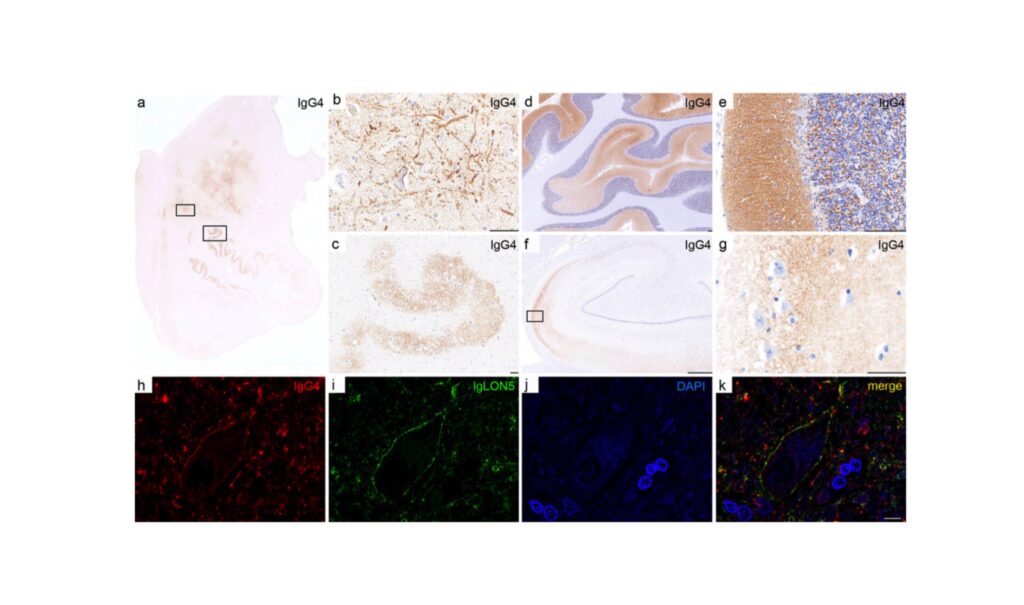
IgLON5 antibodies associated disorder is characterized neuropathologically by the deposition of TAU protein in the hypothalamus and tegmentum of the brainstem. In this cutting-edge work Berger-Sieczkowski et Al., sought to understand whether the chicken or the egg comes first (autoimmunity or neurodegeneration) in IgLON5 disease, studying the neuropathological features in nine patients with this disorder demonstrating that:
- a prominent deposition of anti-IgLON5 IgG4 in the hypothalamus and tegmentum of the brainstem without tau accumulation is a feature of patients with a short disease duration
- a reduced deposition of anti-IgLON5 IgG4 with brainstem neuronal tauopathy is a feature of patients with a long disease duration
- a complement deposition is not found in any patient with igLON5 disease
Overall, this work suggests that anti-IgLON5 antibodies (the autoimmune response) precede TAU pathology (neurodegeneration) in IgLON5 disease. Although, this needs to be demonstrated in future experimental studies, this is an important finding that underscores the importance of early treatment in patients with IgLON5 autoantibodies.

